Tubes Narrow

METHODS

Membrane proteins are some of the most important proteins in biology but they are also some of the most difficult proteins to study. Due to their hydrophobic nature, these proteins tend to aggregate and precipitate in aqueous solutions. For this reason these proteins must be incorporated into detergents or lipids that resemble their native environment. As a result, expression and purification of these proteins can be a daunting task. The methods used in our lab allow us to produce milligram amounts of membrane proteins for our studies.

Expression and Purification of Membrane Proteins

Membrane proteins are incredibly difficult to study due to their hydrophobic nature. Unfortunately, our research requires us to produce milligram amounts of protein for our samples. Recombinant expression of full-length membrane proteins are often avoided due to their lethality to the bacteria during over-expression. Once the protein is expressed it is tremendously difficult to purify the protein due to its propensity to aggregate, even in the presence of detergents or lipids.

Lipid Assemblies

Lipid Assemblies

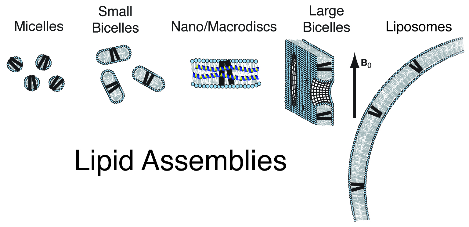
A wide range of detergent and lipid environments have been used for looking at membrane proteins by NMR spectroscopy. By using nanodisks and macrodisks we are able to look at membrane proteins by both solution and solid-state NMR in a membrane-like environment. In order to study the structure, dynamics and function of this protein it will be incorporated into lipid nanodiscs. These lipid assemblies provide the ideal environment for studying these proteins in a bilayer environment. Protein/nanodisc samples will be made into samples for solution and solid-state NMR.

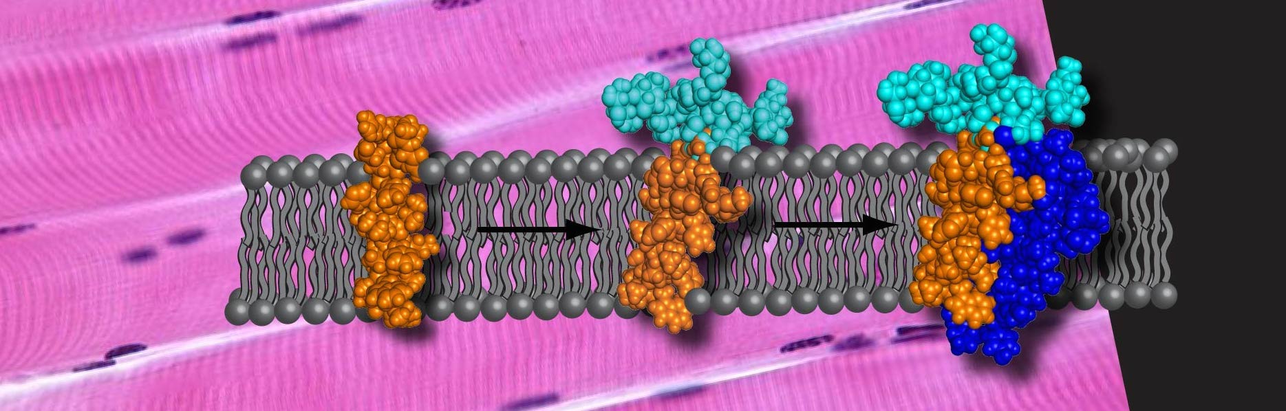
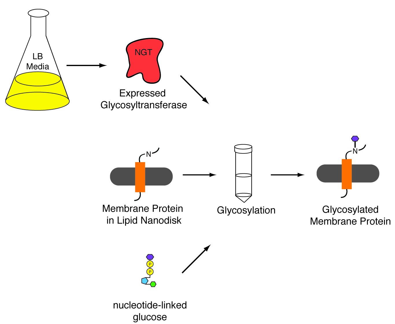
In vitro glycosylation

In order to study our membrane proteins with a saccharide attached we we have to find a way to glycosylate our proteins. Using methods first described by Aebi and co-workers* we will attempt to glycosylate our membrane protein in nanodisks. The nanodisk provides a membrane-like environment that allows the native structure of our membrane protein to be maintained while providing a detergent free system for the glucotransferase to function properly. (*Schwarz, F. et al, JBiolChem, 2011, 286:35267-74)

Protein Glycosylation


NMR

Once these proteins are purified and glycosylated we use Nuclear Magnetic Resonance (NMR) Spectroscopy to determine the structure, dynamics and interactions of these proteins. The method measures the chemical environment around the nuclei of atoms that make up the protein. We can measure bond angles, intramolecular distances and intermolecular distances to develop a 3D model of the protein.

Interaction Studies

The dipolar coupling measurements that acquired can be converted into angle constraints for the structure calculation of our protein structures. A high-resolution 3D structure can be determined using these methods. Other measurements can also be performed including protein-protein and drug-protein interactions. By adding the interacting molecule and measuring the chemical shift perturbations and line-shape changes we are able to determine the precise locations of those interactions. These measurements can be done in the presence and absence of the saccharide to determine the effects that the glycosylation has on these properties as well.


SkeletalMuscle Glycosylation Narrow

EFFORTS

We are interested in looking at proteins that are involved in human disease. By understanding the way they fold, move and interact with other proteins we can better develop therapeutics and treatments to modulate their activity. We are specifically interested in membrane proteins, those proteins that reside in the lipid membranes of cells. We are also looking at how post-translational modification, e.g. glycosylation, affects the physical properties of these proteins.

Glycoproteins

Glycoproteins are a large class of proteins, taking part in nearly every biological process. They participate in the immune system as antibodies and as factors in the major histocompatibilty complex interacting with T cells as part of a the adaptive immune response. They are also involved in white blood cell recognition, cell growth, differentiation, cell-cell interactions and protein folding. Glycoproteins are also indicators for various cancers. A large number of important glycoproteins are integral membrane proteins. They can be found in the lipid bilayers that make up the plasma membrane and the membranes of organelles. In the laboratory of Dr. Cook a combination of solution and solid-state NMR are employed to study the effects of glycosylation on structure, dynamics and the interactions of these important proteins. Understanding these properties is an incredibly important component to the development of treatments of human disease involving glycoproteins

Dystrophin Complex

A large percentage of glycoproteins are membrane proteins. Sarcoglycans are a set of these important proteins. They are responsible for maintaining the integrity of the muscle fiber sarcolemma by connecting the cytoskeleton of the muscle fiber to the extracellular matrix. Very little is known about the structure and dynamics of this set of proteins and of membrane glycoproteins overall. These proteins are not soluble in aqueous environments making them difficult to study by most spectroscopic methods. We have been able to overexpress full-length γ-sarcoglycan in e. coli in an effort to learn more about the structure, dynamics and interactions of this biologically important protein.

Syndecans

Each syndecan has a short cytoplasmic domain, a highly conserved single spanning transmembrane domain, and a large extracellular domain with attachment sites for three to five heparin sulfates or chondroitin sulfates. The syndecan family is involved in the regulation of angiogenesis and cell proliferation, cell-to-cell interaction and cell adhesion through the activation of growth factors. Both syndecan-1 and syndecan-4 have been closely associated with tumor progression. Syndecan-1 was shown to be highly overexpressed on the cell surface in aggressive tumor cells and is associated with poor prognosis. Studies have also demonstrated that the shedding of the syndecan-1 soluble ectodomain inhibits FGF2-induced cell proliferation and is also a potent stimulator for melanoma tumor growth and metastasis. Syndecan-1 is used clinically as a blood plasma tumor marker, through solution NMR spectroscopy. It is thought that the presence of the highly conserved cytoplasmic C1 domain promotes sydecan dimerization. Our computational studies predict that the large ectodomain of syndecan-1 is intrinsically disordered but may adopt a more defined secondary structure upon ligand binding. Despite the significance of these proteins, very little is known about the structure and dynamics of these proteins. To date, only the structure of the cytoplasmic domain of syndecan-4 has been solved. We will use both solution and solid-state NMR to characterize these important proteins and look at the effect that glycosylation has on their properties.

Structure, Dynamics

By determining the structure and dynamics of these proteins we will better understand how they perform their function in biology. Very little is known about membrane glycoproteins and our studies will help other researchers that are working on glycoproteins to better predict their structures and behaviors, leading to a broader knowledge about this important family of proteins.

Protein-Protein and Drug-Protein Interactions

Interactions are another important part of our research. We can use interaction studies to determine how these proteins perform their functions. In certain disease states the blocking of these interactions may be a way to inhibit harmful interactions of proteins that lead to disease. By knowing the precise locations on the protein that are involved in these interactions, drugs can be made and tailored to bind to these sites and block the protein-protein interactions.